
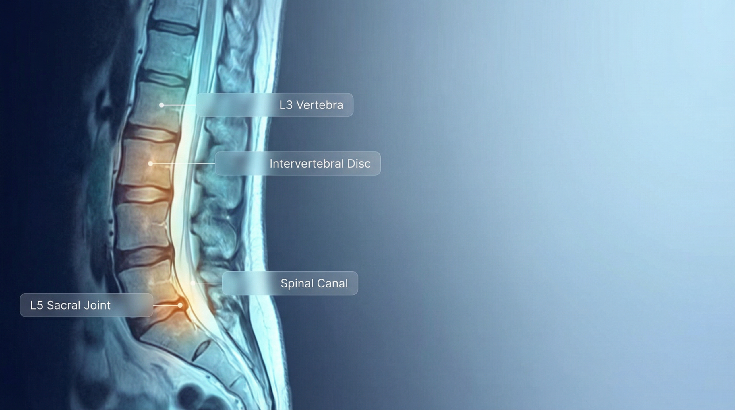
You just got your MRI results back. You open the report and it reads like a foreign language — “broad-based disk protrusion,” “neuroforamina,” “dural sac.” What does any of this actually mean for your back pain?
In this post, we take a real lumbar spine MRI report and show you exactly what ScanExplain does: translate dense medical jargon into a clear, plain-language explanation you can actually understand.
Note: The explanations below are simplified for educational purposes. For personalized medical advice, always consult your doctor.
The Original MRI Report
Here is the full radiology report, exactly as a patient might receive it:
HISTORY: Low back pain.
SEQUENCES: Sagittal T1 and T2 weighted images are supplemented by axial T2 weighted images and by sagittal T2 weighted images obtained in flexion and in extension.
COMMENTS:
Vertebral body signal and vertebral body height are preserved. The overall canal size is unremarkable. The conus medullaris is clear. At L1-L2, L2-L3, L3-L4 and L4-L5 there is normal disk signal and disk height. A few images suggest mild disk bulging to the left at L4-L5.
At L5-S1, there is disk degeneration with loss of disk signal and disk height. There is mild to moderate broad-based disk protrusion impinging upon the anterior aspect of the dural sac and narrowing both neuroforamina and clinical correlation is advised regarding the status of both L5 nerve roots.
Sagittal images obtained in flexion reveal normal range of motion without evidence of instability or pathologic offset. Disk protrusion is somewhat more apparent than in the neutral/sitting position.
Sagittal images obtained in extension reveal normal range of motion without evidence of instability or pathologic offset. Disk protrusion is also somewhat more apparent than in the neutral/sitting position.
IMPRESSION:
1. Moderate diffuse broad-based disk protrusion at the L5-S1 level impinging upon dural sac and narrowing neuroforamina somewhat. Findings are more apparent in flexion and in extension than in the neutral/sitting position.
2. Mild disk bulging L4-L5
If that made your eyes glaze over, you are not alone. Most patients feel the same way. Let’s break it down.
The ScanExplain Translation
Here is how ScanExplain would explain this report to you — section by section, in plain English.
What Was Done
An MRI (magnetic resonance imaging) scan was performed on your lower back — specifically the lumbar spine. The scan took multiple sets of images: some looking at your spine from the side, some in cross-section, and additional images while you bent forward (flexion) and leaned backward (extension). This gives the radiologist a thorough picture of how your spine behaves both at rest and during movement.
The Good News First
The bones of your spine (the vertebrae) look healthy — they have normal shape, size, and signal, meaning there’s no sign of fracture, tumor, or infection. The spinal canal — the tunnel that houses your spinal cord — is a normal size. The bottom tip of your spinal cord (the conus medullaris) also looks clear and healthy.
The disks between vertebrae L1 through L4 all look normal. These are the rubbery cushions that sit between each bone, acting as shock absorbers. At these levels, they have a healthy amount of water content (that’s what “normal signal” means) and they are the right thickness.
The Problem Area: L5-S1 (Bottom of the Spine)
This is the main finding. The disk between your fifth lumbar vertebra (L5) and your sacrum (S1) — the very bottom disk in your lower back — is showing signs of wear:
- Disk degeneration: The disk has lost water content and has gotten thinner. Think of it like a car tire that’s worn down — it’s not as plump and springy as it once was.
- Broad-based disk protrusion: The disk is bulging outward, pushing into the space in front of the dural sac (the protective sleeve around your spinal nerves). “Broad-based” means the bulge spans a wide area rather than poking out at one small point.
- Narrowing of the neuroforamina: The small tunnels where nerves exit the spine on both the left and right sides are getting squeezed. This is significant because the L5 nerve roots pass through here — and if they’re compressed, it can cause pain, tingling, or weakness traveling down into your legs.
A Minor Finding: L4-L5
One level up, there is a mild disk bulge to the left side. This is much less severe than the L5-S1 issue. “Mild bulging” means the disk is slightly pushing outward but is not significantly compressing any nerves or the spinal canal. It’s worth monitoring but is not currently a major concern.
What the Flexion and Extension Images Showed
When you bent forward and leaned backward during the scan, your spine moved normally — no evidence of instability (meaning the vertebrae aren’t slipping out of place). However, the disk protrusion at L5-S1 became more noticeable in both positions compared to when you were sitting upright. This means the bulging disk shifts slightly more during movement, which could explain why your pain changes with certain activities.
Severity Assessment
| Finding | Severity |
|---|---|
| L5-S1 disk degeneration and protrusion | Moderate — the main source of concern |
| Neuroforaminal narrowing (both sides) | Moderate — could be affecting the L5 nerves |
| L4-L5 mild disk bulging | Mild — monitor but low concern for now |
| Spinal stability | Normal — no slippage or instability |
Questions to Ask Your Doctor
Understanding your MRI is just the first step. Here are questions you might want to bring to your next appointment:
- Could the L5-S1 disk protrusion be causing my back pain and any leg symptoms?
- Do I need nerve conduction studies to check if the L5 nerve roots are affected?
- Would physical therapy help, or should we consider other treatment options?
- Should I avoid certain movements or exercises given that the protrusion worsens with flexion and extension?
- How likely is this to progress over time, and how often should I be re-scanned?
From Confusion to Clarity in Seconds
This is exactly what ScanExplain does — takes dense radiology language and turns it into something you can actually understand. No medical degree required. No signup needed.
If you have an MRI, CT, X-ray, or ultrasound report and want to know what it really says, try ScanExplain for free.
Leave a Reply